Comprehensive Cardiac Structure-Function Analysis in Heart Transplantation

Heart transplant (Tx) surgery is a well-established life-saving procedure, but is associated with post-interventional risks such as acute cardiac rejection (ACR), which is one of the leading causes of death in the first year after transplant. Beyond the first year, cardiac allograft vasculopathy (CAV) is the single greatest risk factor for 5-year mortality. Monitoring the patient for post-transplant events is thus paramount. The standard monitoring strategy, however, relies on frequent invasive and costly procedures including endomyocardial biopsies (EMB) and catheter angiography. A reliable non-invasive alternative for the early detection of ACR and CAV would thus be desirable to reduce the need for invasive procedures, improve sensitivity, and reduce cost.
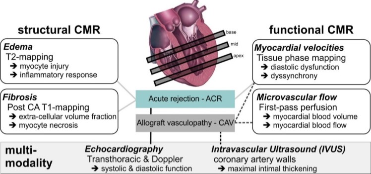
The main goal of this study is to develop a new 15-minute structure-function cardiac MRI protocol for the improved detection of regional abnormalities associated with ACR (edema, fibrosis, dysfunction) and CAV (perfusion, dysfunction). The aim is to help clinicians identify the optimal mixed monitoring strategy, i.e. the optimal combination of multi-modality imaging (structure-function MRI, echo, IVUS) and invasive procedures (EMB, catheter angiography), which provide best outcome (quality adjusted life days) and lowest cost for the individual cardiac transplant patient.
Investigators: Michael Markl (PhD), James Carr (MD), Kai Lin (MD), Robert Gordon (MD), Gordon Hazen (PhD), Dan Lee (MD), Jon Lomasney (MD), Ann Ragin (PhD), Vera Rigolin (MD), Clyde Yancy (MD), Alan Anderson (MD), Roberto Sanari (MD), Julie Blaisdell (MS), Kambiz Ghafourian (MD, MPH), Jonathan Rich (MD)
Funding: National Heart, Lung, And Blood Institute of the NIH (NHLBI)
Publications:
