
Neurovascular 4D Flow
Healthy Controls
4D Flow images of the brain are a rich and multi-dimensional data set. In order to simplify this data while retaining the most important features, we represent the neurovasculature as a flow distribution network graph (FDNG). Here we show (A) time-averaged pathlines derived from the 4D flow velocity information, (B) identification of vessels (yellow) and schematic representation as a FDNG (blue), and (C) a standardized FDNG containing mean and standard deviations of flow values over a group of healthy controls with the same Circle of Willis structure variant.

Brain Arteriovenous Malformation (AVM)
Cerebral arteriovenous malformation (AVM) is a congenital, pathological vascular connection between the arterial and venous cerebral vasculature and has severe complications such as seizure or subarachnoid hemorrhage. An accurate, low-risk hemodynamic assessment across vessel sizes and flow velocities in the cerebral vasculature is needed to improve hemorrhage risk estimates at baseline and throughout staged treatment. We are working to address these needs by developing a non-invasive, non-contrast comprehensive 4D Flow imaging approach to characterize time-resolved 3D blood flow and pressure gradients in AVM. This includes characterizing the hemodynamics of AVMs with 4D flow in order to identify the most clinically relevant metrics and optimizing the 4D flow imaging process to provide that information, from sequence and scan parameters to data post-processing, presentation, and clinical correlation.

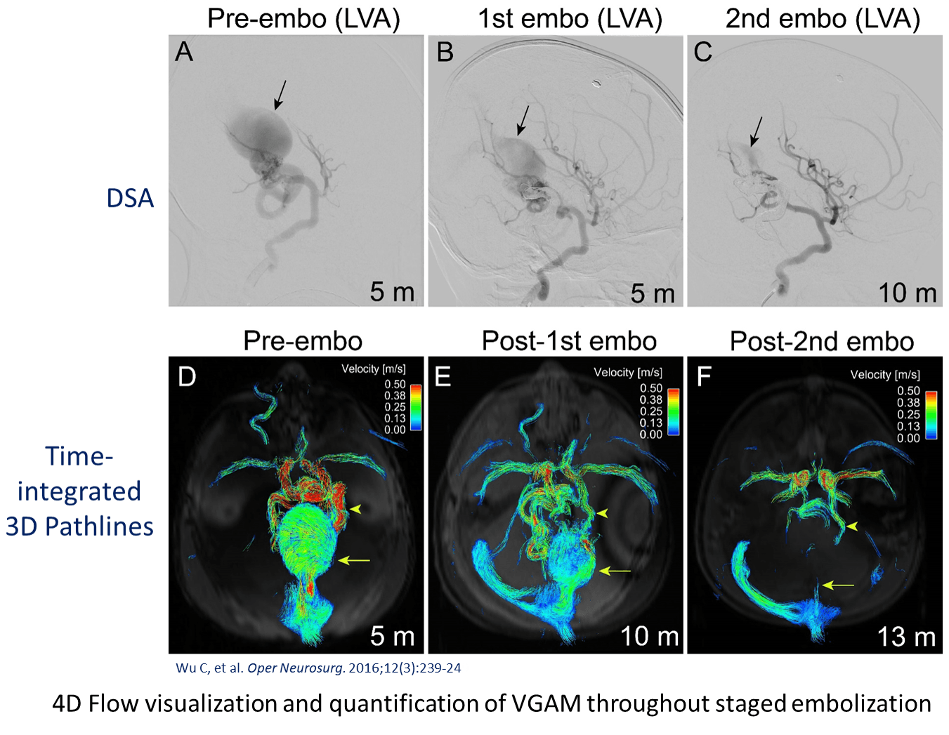
Vein of Galen Aneurysmal Malformation (VGAM)
A Vein of Galen malformation is a specific type of arteriovenous malformation, where a fetal precursor to the Vein of Galen (the vein of Markowski) is supplied directly by arteries and persists at birth as a high-flow shunt between the arterial and venous circulation in the brain. Some VGAM patients are diagnosed in utero or shortly after birth, and others are diagnosed as infants. However, in all cases, the high flow through the shunt creates an increased demand on the heart, leading to heart failure.
Investigation in our lab has focused on characterizing the hemodynamic parameters of these shunts. This work may pave the way for better risk stratification and treatment planning for these patients.
Cerebrovascular Aneurysm

Cerebral aneurysms are a life-threatening condition and too simplified models are currently used to decide on treatment or regular follow-up. Risk of aneurysm growth or rupture is difficult to assess and hemodynamic factors are recently considered as new imaging biomarkers. 4D-flow MRI was employed for the comprehensive in-vivo analysis of hemodynamics and its relationship to size and morphology of different intracranial aneurysms (IA). 4D flow MRI demonstrated the influence of lesion size and morphology on aneurysm hemodynamics suggesting the potential of 4D-flow MRI to assist in the classification of individual aneurysms (1).
We however, that standard 4D flow MRI with single-venc gradients suffers from either velocity aliasing or low velocity to noise ratio. Therefore, we started using dual-venc 4D flow MRI (2) in a new group of patients. We applied and investigated the benefits of using kt-GRAPPA accelerated dual-venc 4D flow MRI in 12 patients who presented with cerebral aneurysm between 4 and 12mm (smallest and largest dimension). When applied to patients with cerebral aneurysms, we could show that dual-venc demonstrated higher sensitivity to slower velocity values and provided less noisy data. Future longitudinal studies and correlation with aneurysm risk factors or aneurysm growth/rupture are required to evaluate the utility of hemodynamic markers for improved risk assessment and therapy planning.
References:
- Schnell S, Ansari SA, Vakil P, Wasielewski M, Carr ML, Hurley MC, et al. Three-dimensional hemodynamics in intracranial aneurysms: Influence of size and morphology. Journal of Magnetic Resonance Imaging. 2014;39:120-131
- Schnell S, Ansari SA, Wu C, Garcia J, Murphy IG, Rahman OA, et al. Accelerated dual-venc 4d flow mri for neurovascular applications. Journal of Magnetic Resonance Imaging. 2017;46:102-114
Intracranial Atherosclerotic Disease (ICAD)
In case of severe ICAD, the stenosis size is below the resolution of the 4D flow MRI. In order to segment these volumes, we used high resolution TOF-MRA to accurately identify and characterize the lesion. This requires co-registration of the images, which can be done automatically in our new analysis tool. This enables the automated identification of vessels and placement of perpendicular planes for quantification.

Funding: German Research Foundation, SIR Research Pilot Grant, RSNA Research Seed Grant, NIH Predoctoral F30 NRSA, AHA Postdoctoral award
Investigators: Susanne Schnell [primary contact] (PhD), Michael Markl (PhD), Can Wu (PhD), Alireza Vali (PhD), Maria Aristova, Ali Shaibani (MD), Sameer Ansari(MD, PhD), Shyam Prabhakaran (MD)
Publications:
Wu C. et al. Age‐Related Changes of Normal Cerebral and Cardiac Blood Flow in Children and Adults Aged 7 Months to 61 Years. JAHA 2016;5:e002657. https://doi.org/10.1161/JAHA.115.002657.
Schnell S, Ansari SA, Wu C, Garcia J, Murphy IG, Rahman OA, Rahsepar AA, Aristova M, Collins JD, Carr JC, Markl M. Accelerated dual-venc 4D flow MRI for neurovascular applications. J Magn Reson Imaging. 2017 Jul; 46(1):102-114. doi: 10.1002/jmri.25595. Epub 2017 Feb 2.
Schnell S, Wu C, Ansari SA. Four-dimensional MRI flow examinations in cerebral and extracerebral vessels – ready for clinical routine? Curr Opin Neurol. 2016 Aug; 29(4):419-28. doi: 10.1097/WCO.0000000000000341.
Markl M, Wu C, Hurley MC, Ansari SA, Carroll TJ, Rahme RJ, Aoun SG, Carr J, Batjer H, Bendok B. Cerebral arteriovenous malformation: Complex 3D hemodynamics and 3D blood flow alterations during staged embolization. JMRI September 2013 https://doi.org/10.1002/jmri.24261
Ansari SA, Schnell S, Carroll T, Vakil P, Hurley MC, Wu C, Carr J, Bendok BR, Batjer H, Markl M. Intracranial 4D flow MRI: toward individualized assessment of arteriovenous malformation hemodynamics and treatment-induced changes. AJNR Am J Neuroradiol. 2013 Oct;34(10):1922-8. doi: 10.3174/ajnr.A3537. Epub 2013 May 2.Wu, S.A. Ansari, A.R. Honarmand, P. Vakil, M.C. Hurley, B.R. Bendok, J. Carr, T.J. Carroll and M. Markl. Evaluation of 4D Vascular Flow and Tissue Perfusion in Cerebral Arteriovenous Malformations: Influence of Spetzler-Martin Grade, Clinical Presentation, and AVM Risk Factors. American Journal of Neuroradiology June 2015, 36 (6) 1142-1149; DOI: https://doi.org/10.3174/ajnr.A4259
Wu C. et al. Complex Alterations of Intracranial 4-Dimensional Hemodynamics in Vein of Galen Aneurysmal Malformations During Staged Endovascular Embolization. Operative Neurosurgery, Volume 12, Issue 3, 1 September 2016, Pages 239–249, https://doi.org/10.1227/NEU.0000000000001137.
Schnell S, Ansari SA, Vakil P, Wasielewski M, Carr ML, Hurley MC, Bendok BR, Batjer H, Carroll TJ, Carr J, Markl M. Three-dimensional hemodynamics in intracranial aneurysms: influence of size and morphology. J Magn Reson Imaging. 2014 Jan;39(1):120-31. doi: 10.1002/jmri.24110. Epub 2013 Oct 22.
Wu C, Schnell S, Vakil P, Honarmand AR, Ansari SA, Carr J, Markl M, Prabhakaran S. In Vivo Assessment of the Impact of Regional Intracranial Atherosclerotic Lesions on Brain Arterial 3D Hemodynamics. AJNR Am J Neuroradiol. 2017 Mar;38(3):515-522. doi: 10.3174/ajnr.A5051. Epub 2017 Jan 5.